Útskýring á HD og ED
Mjaðma og olnbogamyndatökur fyrir ræktun er hægt að senda bæði til SKK /svíþjóðar og OFA/bandaríkin. Hægt er að senda röntgen myndir af 1 árs hundi til aflestrar og fá staðfestar/gildar niðurstöður hjá SKK en hjá OFA þarf hundurinn að vera 2ja ára. SKK-Svíðþjóð Ef á að senda myndir til SKK þá þarf eigandi að kaupa aflestur fyrirfram fyrir hundinn svo að dýralæknir geti sent myndir til SKK til aflestrar það er gert hér https://www.mittskk.se/hd-ed-reading-fee-dogs-from-iceland.html einnig þarf að fylla út umsókn og senda umsóknina til [email protected] OFA-Bandaríkin Röntgen myndir sendar til OFA þá er hægt að fylla út umsókn hér beint inn á síðunni þeirra eða fá útprentaða umsókn hér eða biðja dýralækninn um það. Þegar mætt er til dýralæknis þarf að hafa ættbókina frá HRFÍ eða vera búin að senda mynd af ættbókinni til dýralæknisns.
HD - Mjaðmir Niðurstaða og þýðir Fri - A Excellent - A1 Good - A2 Fair - B Borderline*- Óstaðfest niðurstaða, þarf að endurtaka myndatöku eftir 6.mán. Mild / Svak / Mild (US) - C Moderat / Middels / Moderate (US) - D Severe / Sterk / Severe (US) - E * Borderline: there is no clear cut consensus between the radiologists to place the hip into a given category of normal or dysplastic. There is usually more incongruency present than what occurs in the minor amount found in a fair but there are no arthritic changes present that definitively diagnose the hip joint being dysplastic. There also may be a bony projection present on any of the areas of the hip anatomy illustrated above that can not accurately be assessed as being an abnormal arthritic change or as a normal anatomic variant for that individual dog. To increase the accuracy of a correct diagnosis, it is recommended to repeat the radiographs at a later date (usually 6 months). This allows the radiologist to compare the initial film with the most recent film over a given time period and assess for progressive arthritic changes that would be expected if the dog was truly dysplastic. Most dogs with this grade (over 50%) show no change in hip conformation over time and receive a normal hip rating; usually a fair hip phenotype.
ED - Olnbogi Niðurstaða og þýðir 0 / Fri - A 1 / Mild / Svak - C 2 / Moderat / Middels - D 3 / Severe / Sterk - E
+++++++++++++++++++++++++++++++++++++++++++++ What Do Hip Grades Mean?The phenotypic evaluation of hips done by the OFA falls into seven different categories. Those categories are Normal (Excellent, Good, Fair), Borderline, and Dysplastic (Mild, Moderate, Severe). Once each of the radiologists classifies the hip into one of the 7 phenotypes above, the final hip grade is decided by a consensus of the 3 independent outside evaluations. Examples would be:
The hip grades of Excellent, Good and Fair are within normal limits and are given OFA numbers. This information is accepted by AKC on dogs with permanent identification (tattoo, microchip) and is in the public domain. Radiographs of Borderline, Mild, Moderate and Severely dysplastic hip grades are reviewed by the OFA radiologist and a radiographic report is generated documenting the abnormal radiographic findings. Unless the owner has chosen the open database, dysplastic hip grades are not in the public domain. ++++++++++++++++++++++++++++++++++++++++++++++++++++++++++++++
ExcellentExcellent: this classification is assigned for superior conformation in comparison to other animals of the same age and breed. There is a deep seated ball (femoral head) which fits tightly into a well-formed socket (acetabulum) with minimal joint space. There is almost complete coverage of the socket over the ball +++++++++++++++++++++++++++++++++++++++++++++++++++++ GoodGood: slightly less than superior but a well-formed congruent hip joint is visualized. The ball fits well into the socket and good coverage is present. ++++++++++++++++++++++++++++++++++++++++++++++++++++++++++ FairFair: Assigned where minor irregularities in the hip joint exist. The hip joint is wider than a good hip phenotype. This is due to the ball slightly slipping out of the socket causing a minor degree of joint incongruency. There may also be slight inward deviation of the weight-bearing surface of the socket (dorsal acetabular rim) causing the socket to appear slightly shallow. This can be a normal finding in some breeds however, such as the Chinese Shar Pei, Chow Chow, and Poodle. ++++++++++++++++++++++++++++++++++++++++++++++ BorderlineBorderline: there is no clear cut consensus between the radiologists to place the hip into a given category of normal or dysplastic. There is usually more incongruency present than what occurs in the minor amount found in a fair but there are no arthritic changes present that definitively diagnose the hip joint being dysplastic. There also may be a bony projection present on any of the areas of the hip anatomy illustrated above that can not accurately be assessed as being an abnormal arthritic change or as a normal anatomic variant for that individual dog. To increase the accuracy of a correct diagnosis, it is recommended to repeat the radiographs at a later date (usually 6 months). This allows the radiologist to compare the initial film with the most recent film over a given time period and assess for progressive arthritic changes that would be expected if the dog was truly dysplastic. Most dogs with this grade (over 50%) show no change in hip conformation over time and receive a normal hip rating; usually a fair hip phenotype. ++++++++++++++++++++++++++++++++++++++++++++++++++++ MildMild Hip Dysplasia: there is significant subluxation present where the ball is partially out of the socket causing an incongruent increased joint space. The socket is usually shallow only partially covering the ball. There are usually no arthritic changes present with this classification and if the dog is young (24 to 30 months of age), there is an option to resubmit an radiograph when the dog is older so it can be reevaluated a second time. Most dogs will remain dysplastic showing progression of the disease with early arthritic changes. Since HD is a chronic, progressive disease, the older the dog, the more accurate the diagnosis of HD (or lack of HD). +++++++++++++++++++++++++++++++++++++++++++++++++++++ ModerateModerate Hip Dysplasia: there is significant subluxation present where the ball is barely seated into a shallow socket causing joint incongruency. There are secondary arthritic bone changes usually along the femoral neck and head (termed remodeling), acetabular rim changes (termed osteophytes or bone spurs) and various degrees of trabecular bone pattern changes called sclerosis. Once arthritis is reported, there is only continued progression of arthritis over time. ++++++++++++++++++++++++++++++++++++++++++++++++++++ SevereSevere Hip Dysplasia: assigned where radiographic evidence of marked dysplasia exists. There is significant subluxation present where the ball is partly or completely out of a shallow socket. Like moderate HD, there are also large amounts of secondary arthritic bone changes along the femoral neck and head, acetabular rim changes and large amounts of abnormal bone pattern changes ++++++++++++++++++++++++++++++++++++++++++++++++++++ The Three Faces of Elbow DysplasiaElbow dysplasia is a general term used to identify an inherited polygenic disease in the elbow. Three specific etiologies make up this disease and they can occur independently or in conjunction with one another. These etiologies include:
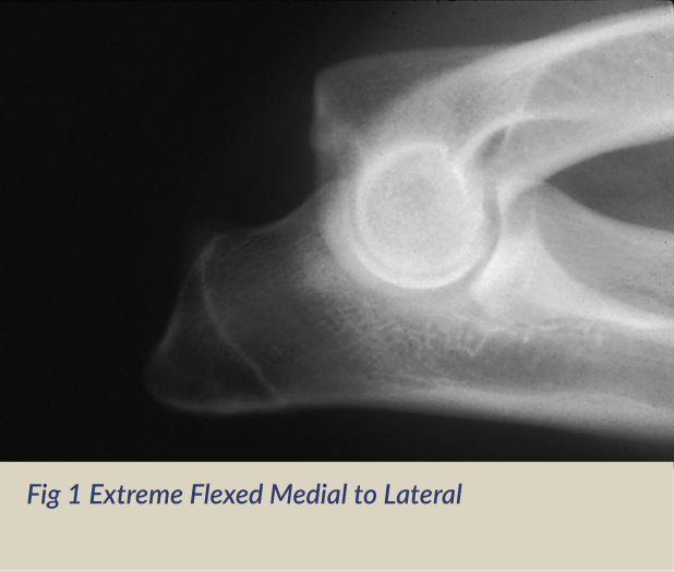
Studies have shown the inherited polygenic traits causing these etiologies are independent of one another. Clinical signs involve lameness which may remain subtle for long periods of time. No one can predict at what age lameness will occur in a dog due to a large number of genetic and environmental factors such as degree of severity of changes, rate of weight gain, amount of exercise, etc.. Subtle changes in gait may be characterized by excessive inward deviation of the paw which raises the outside of the paw so that it receives less weight and distributes more mechanical weight on the outside (lateral) aspect of the elbow joint away from the lesions located on the inside of the joint. Range of motion in the elbow is also decreased. Elbow Dysplasia GradesElbow dysplasia in dogs has multiple inherited etiologies which may occur singularly or in combination. These etiologies include fragmented medial coronoid (FCP) of the ulna, osteochondritis of the medial humeral condyle and ununited anconeal process (UAP). The most sensitive view used to diagnose secondary degenerative changes in the elbow joint is an extreme flexed mediolateral view of the elbow which is required by the OFA and recommended by the International Elbow Working Group. Veterinary radiologists are most interested in the appearance of the anconeal process of the ulna. When there is instability of the elbow joint due to elbow dysplasia in a dog, one of the most sensitive radiographic findings is new bone proliferation (osteophytes) on the anconeal process of the ulna associated with secondary developmental degenerative joint disease. Bone proliferation can be very subtle to visualize in some dogs. Other arthritic findings such as sclerosis in the area of the trochlear notch of the ulna and bone spurs at joint edges are also reported. If fragmentation of the medial coronoid only involves the cartilage, it may not be seen radiographically but occasionally if the bone is also fragmented, it can be visualized as a separate calcific opacity superimposed over the radius.    Explanation of Elbow GradesFor elbow dysplasia evaluations for dogs, there are no grades for a radiographically normal elbow. The only grades involved are for abnormal elbows with radiographic changes associated with secondary degenerative joint disease. Like the hip certification, the OFA will not certify a normal elbow until the dog is 2 years of age. The OFA also accepts preliminary elbow radiographs. To date, there are no long-term studies for preliminary elbow examinations like there are for hips; however, preliminary screening for elbows along with hips can also provide valuable information to the breeder. Grade I Elbow Dysplasia: Minimal bone change along anconeal process of ulna (less than 2mm). Radiographic Elbow ViewsThe International Elbow Working Group (IEWG), a consortium of experts from around the world was founded in 1989 to lower the incidence of elbow dysplasia by coordinating worldwide efforts. The diagnosis of elbow dysplasia is based on the presence of degenerative joint disease/osteoarthritis which results from an ununited anconeal process, osteochondrosis or a fragmented medial coronoid process. The OFA started its elbow database in 1990 using a modified protocol of the IEWG. For cost effectiveness and mass screening, the OFA requires the extreme flexed medial to lateral view (Fig 1) of each elbow. Inclusion of additional views such as the neutral medial to lateral (Fig 2), cranial caudal (Fig 3) or preferably the cranial caudal 10-15 degree lateral to medial oblique (Fig 4) can also be submitted and will be evaluated. In instances where the owner is requesting a re-evaluation of the elbows or if the dog is experiencing lameness it is recommended that the cranial caudal 10 to 15 degree lateral to medial oblique view accompany the required extreme flexed medial-lateral view.
|
Schafer Nafn: KRISTJANAFarsími: 790-6868Tölvupóstfang: [email protected]Um: Schafer ræktun - Þýskur fjárhundurSýningar- Þjálfun kl 19 Það er í dag! Ice Tindra ganga kl 13 atburður liðinn í 11 daga Winter Wonderland sýning eftir 16 daga Alþjóðlegsýning HRFÍ 20.-21.feb 2027 eftir 3 mánuði 9 daga Orivet heilsutest atburður liðinn í 3 ár 2 mánuði 6 daga Belcando hundafóður atburður liðinn í 7 ár 7 mánuði 10 daga DNA-test atburður liðinn í 5 ár 7 mánuði 15 daga Tenglar
|